6.1 The first central station: the cochlear nucleus
The auditory nerve from each cochlea enters the brainstem at the cochlear nucleus (CN), which is divided into three subdivisions: the anteroventral CN (AVCN), the posteroventral CN (PVCN), and the dorsal CN (DCN). Each auditory-nerve fiber bifurcates as it enters the CN, sending one branch to AVCN and the other to PVCN/DCN.
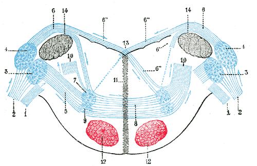
Each subdivision has its own characteristic cell types and output projections. AVCN contains the famous bushy cells, which receive a small number of giant endbulb of Held synapses from auditory-nerve fibers. The endbulb is one of the largest synapses in the brain; a single auditory-nerve fiber forms a calyx that engulfs much of the bushy cell’s soma, and a single presynaptic spike reliably triggers a postsynaptic spike with extraordinary temporal fidelity. The bushy cells are designed to preserve timing, not transform it — they pass spike trains forward with timing jitter on the order of tens of microseconds.
The DCN is more complex, containing pyramidal cells that perform spectral analysis (likely involved in elevation cues), giant cells that integrate multimodal inputs, and a layered organization that hints at cortex-like processing.
For the purposes of localization, the structures we care about are downstream of the AVCN bushy cells.